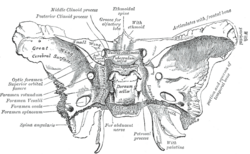
棘孔
| 棘孔 | |
|---|---|
 蝶骨內面觀,棘孔標示為左下方往上數第二個 | |
 顱底內面觀,蝶骨標為黃色,棘孔可見於右下方 | |
| 標識字符 | |
| 拉丁文 | Foramen spinosum |
| TA98 | A02.1.05.038 |
| TA2 | 624 |
| FMA | FMA:53156 |
| 格雷氏 | p.150 |
| 《骨骼解剖學術語》 [在維基數據上編輯] | |
棘孔(Foramen spinosum)是人類顱底的一對孔洞,位於蝶骨棘的前方、卵圓孔的側邊,兩側各一。通過棘孔的構造有中腦膜動脈、中腦膜靜脈和下頷神經腦膜支。
棘孔常在神經外科手術中作為定位點,可藉由相對位置協助辨識其他顱底孔洞。18世紀時,賈柯柏·貝尼格納斯·溫斯洛首先描述了棘孔。
結構
[編輯]棘孔位於中顱窩內,是一個貫穿蝶骨的孔洞,[1][2]:771,和卵圓孔並列為蝶骨大翼上唯二的兩對孔洞,卵圓孔則位於棘孔的前內側[2]:776。棘孔的後內側為蝶骨棘,外側為下頷窩[2]:873,後方為耳咽管[1]。
變異
[編輯]棘孔的大小和位置因人而異。沒有棘孔的人非常罕見,且通常只有單側缺失,在此情形下,中腦膜動脈由卵圓孔進入顱腔[3]。棘孔也可能邊緣不完整,估計半數的人有此現象;相反地,只有很少數的人(小於1%)有兩對棘孔,這種些人也常有兩對中腦膜動脈[1][3]。棘孔通常從蝶骨棘尖端或內側表面(靠體軸中心處)穿過蝶骨[1]。
發育
[編輯]新生兒的棘孔長約2.25毫米,寬1.05毫米;成人棘孔則長約2.56毫米,寬2.1毫米[4],平均半徑2.63毫米[5]。在一份研究棘孔、圓孔和卵圓孔發育的研究中,棘孔最早在出生後八個月、最遲在七歲前形成完整的環狀構造,多數人最終會發育成圓形[5]。蝶顎韌帶由咽弓發育而來,通常附着在蝶骨棘上,因此可能在棘孔邊緣發現這條韌帶[1][6]。
其它動物
[編輯]其他人科動物棘孔的位置不在蝶骨,而是在顳骨上,例如顳骨鱗狀部或顳骨和蝶骨交界的蝶鱗縫,但也可能沒有棘孔[1][7]。
功能
[編輯]棘孔使中腦膜動脈、中腦膜靜脈和下頜神經腦膜支能通過顱骨,進入顱腔[1][2]:763。
臨床重要性
[編輯]棘孔常因為其特殊的位置而在神經外科手術中作為定位點,用來判斷其他顱底孔洞(包括圓孔和卵圓孔)、下頜神經和三叉神經節的位置。顱內創傷手術的止血也可能涉及棘孔。[1]
歷史
[編輯]18世紀時,丹麥解剖學家賈克柏·溫斯洛首次描述了棘孔,由於這個孔洞和蝶骨大翼的棘突位置相近,因此被命名為棘孔。然而,棘孔的拉丁文「foramen spinosum」在命名時使用了錯誤的詞形變化,因此字面上的意思變成了「有很多棘的孔洞」;正確的詞形變化應為「foramen spinae」,但反而比較少用[1]。
參見
[編輯]本條目使用了部分解剖術語。
參考資料
[編輯]本條目包含來自屬於公共領域版本的《格雷氏解剖學》之內容,而其中有些資訊可能已經過時。
- ^ 1.0 1.1 1.2 1.3 1.4 1.5 1.6 1.7 1.8 Krayenbühl, Niklaus; Isolan, Gustavo Rassier; Al-Mefty, Ossama. The foramen spinosum: a landmark in middle fossa surgery. Neurosurgical Review. 2 August 2008, 31 (4): 397–402. doi:10.1007/s10143-008-0152-6.
- ^ 2.0 2.1 2.2 2.3 Drake, Richard L.; Vogl, Wayne; Tibbitts, Adam W.M. Mitchell; illustrations by Richard; Richardson, Paul. Gray's anatomy for students. Philadelphia: Elsevier/Churchill Livingstone. 2005. ISBN 978-0-8089-2306-0.
- ^ 3.0 3.1 Illustrated Encyclopedia of Human Anatomic Variation: Opus V: Skeletal Systems: Cranium – Sphenoid Bone. Illustrated Encyclopedia of Human Anatomic Variation. [2006-04-10]. (原始內容存檔於2006-05-19).
- ^ Lang J, Maier R, Schafhauser O. Postnatal enlargement of the foramina rotundum, ovale et spinosum and their topographical changes. Anatomischer Anzeiger. 1984, 156 (5): 351–87. PMID 6486466.
- ^ 5.0 5.1 Yanagi S. Developmental studies on the foramen rotundum, foramen ovale and foramen spinosum of the human sphenoid bone. The Hokkaido Journal of Medical Science. 1987, 62 (3): 485–96. PMID 3610040.
- ^ Ort, Bruce Ian Bogart, Victoria. Elsevier's integrated anatomy and embryology. Philadelphia, Pa.: Elsevier Saunders. 2007: Elsevier’s Integrated Anatomy and Embryology. ISBN 978-1-4160-3165-9.
- ^ Braga, J.; Crubézy, E.; Elyaqtine, M. The posterior border of the sphenoid greater wing and its phylogenetic usefulness in human evolution. American Journal of Physical Anthropology. 1998, 107 (4): 387–399. PMID 9859876. doi:10.1002/(SICI)1096-8644(199812)107:4<387::AID-AJPA2>3.0.CO;2-Y.
外部連結
[編輯]- 人體解剖學在線(Human Anatomy Online)網站上的相關圖片:22:os-0902 – "Osteology of the Skull: Internal Surface of Skull"
- Anatomy figure: 27:02-03 at Human Anatomy Online, SUNY Downstate Medical Center – "Schematic view of key landmarks of the infratemporal fossa."
- Anatomy of the Skull – 27. Foramen spinosum (頁面存檔備份,存於互聯網檔案館)
